Service

We Combine In-vitro-Grown Human Organoids with Deep-Learning AI Analysis Technology.

Organoid Live Imaging Analysis

Cardiac Organoid Live Imaging Analysis

Using live brightfield and fluorescence imaging, we quantify a range of cardiac functional parameters in cardiac organoids.

심장 오가노이드 Live 영상 분석

Customized Organoid Imaging Analysis

We perform tailored functional analyses on the imaging data you provide.

  • Beating rate and ejection fraction measurement

  • Electrical signal propagation analysis and optical mapping

  • Real-time calcium transient and conduction velocity analysis

  • Contractile motion pattern analysis

  • Other customized analyses

  • *Please contact us in advance to discuss your analysis requirements.

    Contact Us

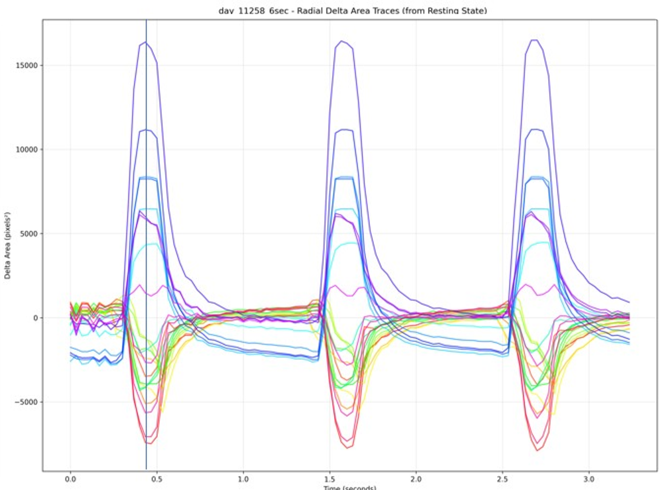
Cardiac Organoid Imaging Parameters

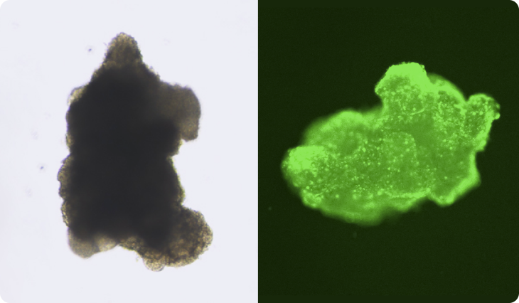
Brightfield imaging
광학 영상 분석 예시
Optical
No. Parameter Description
01 Beat Rate (bpm) Beats per minute across the entire field of view
02 Beat Rate Variation Variation in inter-beat intervals across the entire field of view
03 Velocity (pixels/sec) Contraction velocity
04 Contraction Displacement (pixels) Magnitude of contractile displacement
05 Peak Height Variation Variation in peak amplitude between beats
06 Duration (75%) (sec) Contraction duration measured at 75% of peak amplitude
07 Duration (50%) (sec) Contraction duration measured at 50% of peak amplitude
08 Contraction Time (sec) Time from full relaxation to peak contraction
09 Relaxation Time (sec) Time from peak contraction to full relaxation
10 Diastolic Dimension (if cavity present) Internal cavity diameter at diastole
11 Systolic Dimension (if cavity present) Internal cavity diameter at systole
12 Fractional Shortening (if cavity present) Fractional shortening of the cavity diameter
13 Ejection Fraction (if cavity present) Ejection fraction (%)
14 Cardiac Wall Thickness (if cavity present) Wall thickness at diastole and systole
Calcium transient (fluorescence) imaging
칼슘변이 형광 영상 분석 예시
Calcium variation (fluorescence)
No. Parameter Description
01 Beat Rate (bpm) Beats per minute across the entire field of view
02 Beat Rate Variation Variation in inter-beat intervals across the entire field of view
03 Velocity (pixels/sec) Contraction velocity
04 Contraction Displacement (pixels) Magnitude of contractile displacement
05 Peak Height Variation Variation in peak amplitude between beats
06 Duration (75%) (sec) Contraction duration measured at 75% of peak amplitude
07 Duration (50%) (sec) Contraction duration measured at 50% of peak amplitude
08 Contraction Time (sec) Time from full relaxation to peak contraction
09 Relaxation Time (sec) Time from peak contraction to full relaxation
10 Calcium Fluorescence Change, F/F₀ Magnitude Magnitude of the calcium transient
11 Rise Time Time to peak calcium concentration
12 CTD90 Calcium transient duration at 90% decay
13 T50% Time to 50% decay of the calcium transient
14 D/R Ratio Ratio of calcium release time to calcium influx time
15 Decay Constant (T) Time constant of the calcium decay curve
  • POINT 01
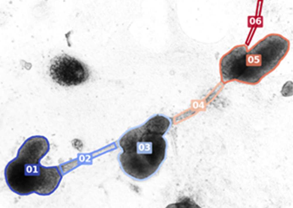
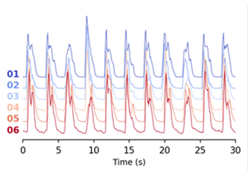
    Electrical signal propagation in 2D cardiac organoids
    2D cardiac organoid electrical signal transmission 1
    2D cardiac organoid electrical signal transmission 2
  • POINT 02
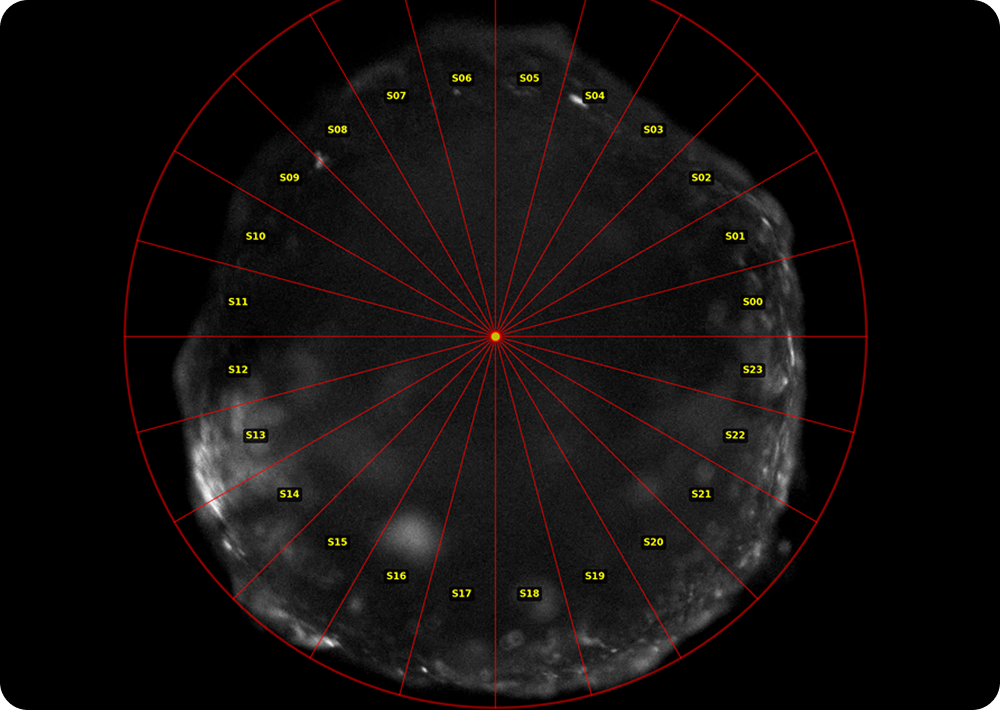
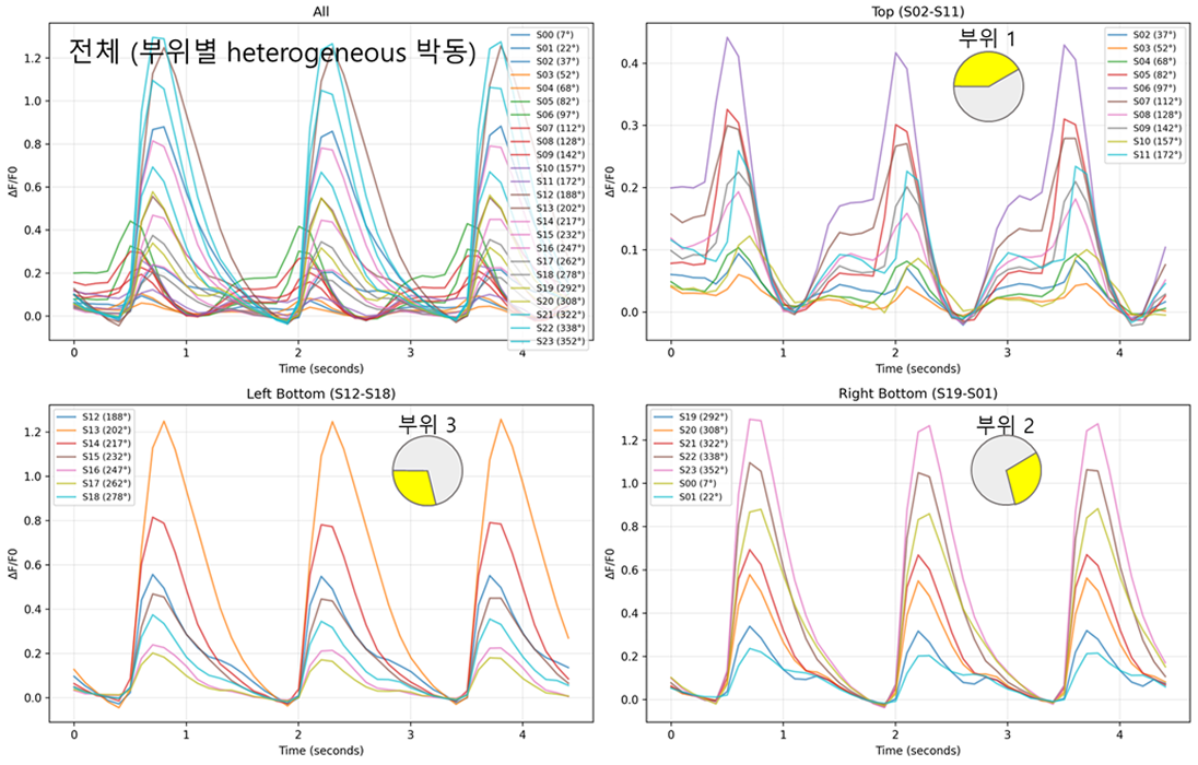
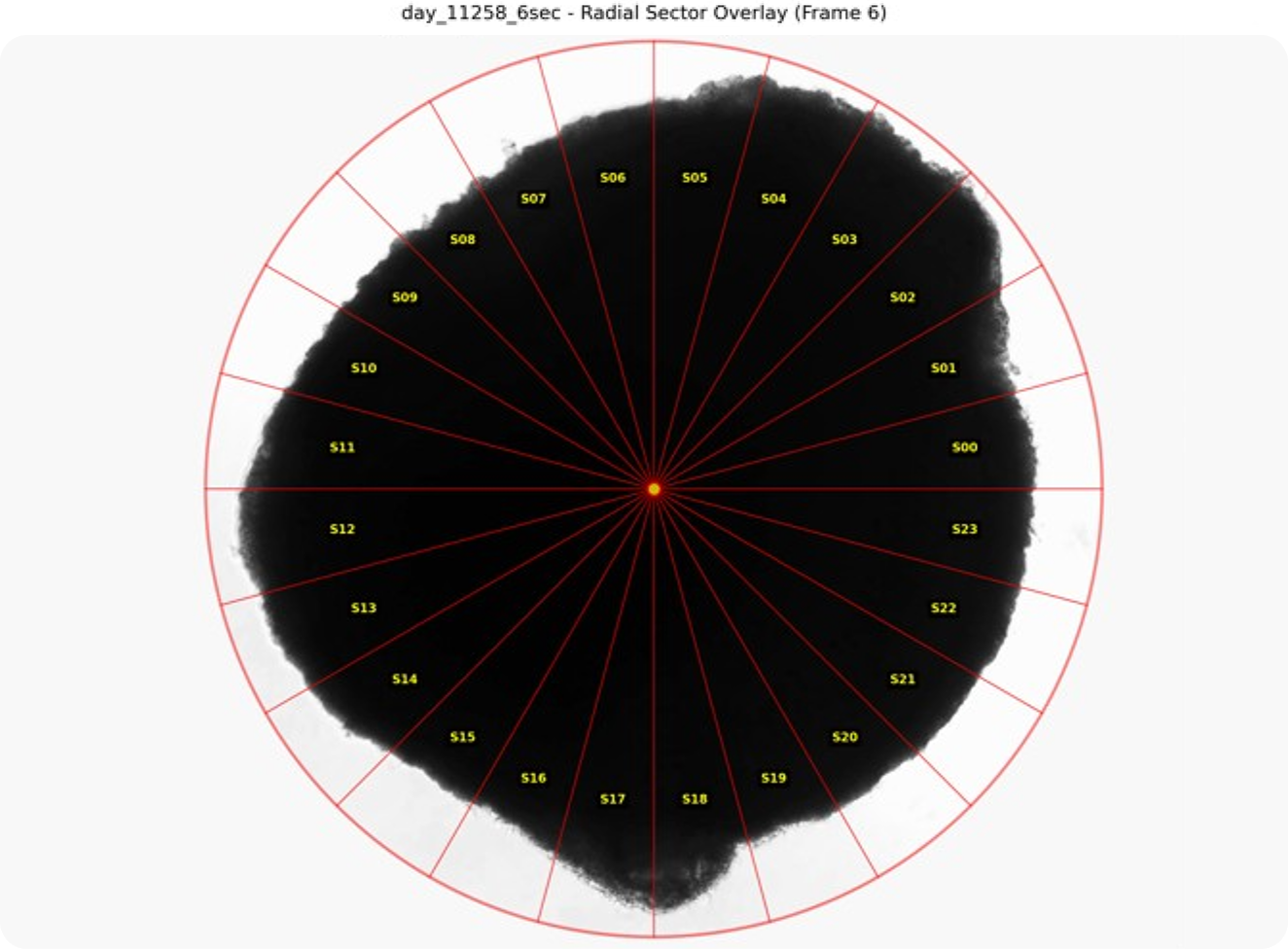
    Assessment of regional beating synchrony within a single organoid using calcium transient fluorescence imaging
    Evaluation of pulsation consistency by region in a single organoid
							using calcium variation fluorescence imaging 1
    Evaluation of pulsation consistency by region in a single organoid
							using calcium variation fluorescence imaging 2
  • POINT 03
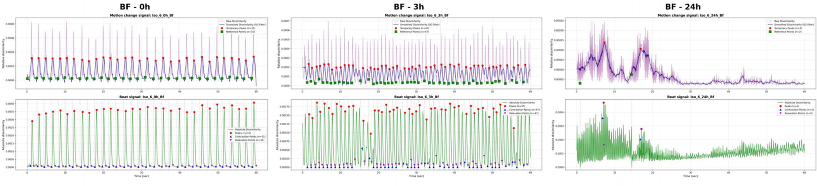
    Time-course analysis of beating rate variability after drug treatment
    Hourly heart rate variability analysis after drug administration